Search the Community
Showing results for tags 'ABO/RH'.
Found 3 results
-
Human versus monoclonal reagents
Silly question...,but I'd really like to know,what is the difference in choosing between human and monoclonal reagents for ABO/D testing? Is it a first choice from these two?Is it the price? I work with Bio-Rad reagents and I know they have cards with human reagents but also identical cards with monoclonal reagents.Our lab use only monoclonal ones.
-
Anti-D Testing Mystery
Hi All, So I would like to present a scenario that happened to me and get your input. I received a specimen from the ED for ABO/Rh testing on a young female (she had a miscarriage, which at the time I was not aware). We use the BD Pink (EDTA) blood bank tubes for all of our blood bank testing, this particular sample was about a little more than 1/4 of the way full (yes not the best sample, learned my lesson with this case) - there were no visible clots in the tube or in the cell suspension I made for testing (testing was done fairly quickly since it was only an ABO/Rh and we use a STATSpin centrifuge so I had results out within 20mins of it being collected). These were my initial reactions at time of testing: Anti-A: 0 Anti-B: 4+ Anti-D: w1+ D Control: 0 A1 Cells: 4+ B Cells: 0 Du: 3+ Du Control: 0 CCC: 3+ So of course my interpretation was B Positive, which was reported. (We use the monoclonal Anti-D) All of our samples are retested by another technologist if we have no previous history on the patient. The samples sit at room temperature until they are tested and this was one was retested roughly 10 hours after my initial testing, these are the results: Anti-A: 0 Anti-B: 4+ Anti-D: 0 D Control: 0 A1 Cells: 4+ B Cells: 0 Du: 0 CCC: 3+ Du Control: 0 CCC: 3+ Interpretation: B Negative It was tested 2 more times by two other techs later that morning and the report corrected and the patient had to be called back to get Rhogam injection. So of course and event report was initiated at that point for root cause analysis. I was approached 3 days later after knowing nothing about what happened and told that I "miss-typed" a patient sample. After reviewing the work card I of course said No I didn't because I actually remember working on this particular sample due to the fact that the D got stronger at AHG phase. I was extremely puzzled by the results and pulled the sample at this point had been in the refrigerator for 3 days and found that the same had numerous large clots and lots of visible small clots in my cell suspension (however none of the previous techs noted or expressed this to my director). I performed (3 days later) a forward and reverse typing (B Neg), DAT, and IAT. The DAT (IgG and Poly) - Both negative (very sticky microscopically) and IAT in Gel was completely negative. So at this point it was very perplexing as to what happened with the Anti-D. Of course everyone my boss talked to said that this was impossible for the Anti-D to just disappear (I was not inferring that it disappeared to her) and I must have done something wrong, which really aggravates me to use the word "impossible" in medicine. She said it was more logical that I mixed the control tube and the Anti-D tube at the AHG phase when I read the tubes which makes absolutely no sense to me (if the results were reversed from the results I got at immediate spin, then yes that would make sense and I would have questioned the results and started over). OK, so here is my theory as to be the possible cause for this particular scenario has to do with a sub-optimal sample that was in the process of clotting at the time of initial testing - however I can only assume this theory and I know that it didn't interfere with the Anti-A or Control on the forward type, but these are all different antiseras with different blends of antibodies/proteins, etc.. I am thinking that since I didn't see or detect clots when I tested the EDTA sample the first time it is possible that something during the clotting process potentially interfered with the Anti-D causing it to be a false positive. Since the specimen wasn't tested again until 10 hours later when the clotting process was definitely complete there is no way to prove this, unless someone where to have retested it immediately after I performed the first test. We've seen cold autoantibodies disappear after sitting at room temperature due to autoadsorption, who is to say that since the clotting process was complete whatever was causing the interference may have been gone 10 hours later when the full clots formed. My boss refuses to even think this is remotely a possible and I had to have made an error, she said we use to use plain red clotted tubes all the time for blood bank and never had any problems (don't think she is getting that these tubes have no additive and normally the clotting process was a lot faster in plain red tubes with no additive. Then again she also couldn't understand how a clot would get stronger overtime (I wanted to bang my head on the desk with that remark). If I made an error or potentially made an error I would definitely own up to it, but in this instance her suggestion that I switched the test tubes around and reading the control as the patient makes no sense what so ever. Has anyone every encountered anything like this before and what is your thoughts on the potential reason for the various reactions between a 10 hour period? Thanks, -TxLabGuy82
-
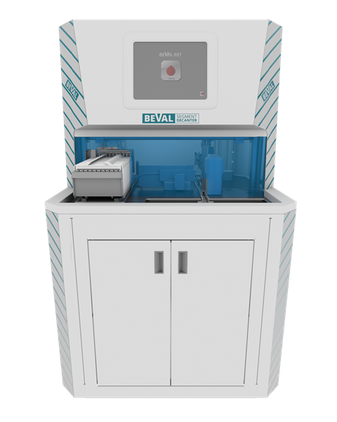
Automated segment decanter for cross match testing
In South Africa it is legislated that the ABO/Rh be confirmed on the segment from the pack before transfusion. When the blood bank changed to an automated crossmatch method, segment testing remained manual due to the cost of automated grouping. The cost of testing the ABO/Rh on the Olympus PK7300 proved to be significantly lower than other methods like Autovue etc. While there were substantial benefits new problems emerged. The tediousness of the task of cutting the segments manually to decant the blood into the test tubes (800-1000 per staff member), as well as manually comparing the bar-code numbers. We have now developed an automated segment cutter. The segment cutter prints the bar-code on the test tube, cuts the segments, decants the blood and adds saline. The racks with test tubes can now be placed directly onto the Olympus for analyzing. The decanter can process up to 300 segments per hour and sends the information to the LIS. Now my questions are the following: • Are the cross matching processes the same globally, i.e. cross match on segment not on specimen? • How common is it that the blood banks do all testing in bulk and send final product to hospital? I apologize in advance if I have some of the terminology wrong. I designed the machine and my blood testing knowledge is limited. Any information would be greatly appreciated. Thanks in advance.
